上海埃飞科技
Worldwide Technology(S. H)上海埃飞科技
Worldwide Technology(S. H)产品展示

Small animal fundus mirror imaging system
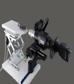
VivoTM small animal ophthalmoscope is customized for your current and future research. Versatility makes our system flexible and valuable. Our basic imaging system provides the function of fluorescein angiography.
VivoTM small animal ophthalmoscope is customized for your current and future research. Versatility makes our system flexible and valuable. Our basic imaging system provides the function of fluorescein angiography.
When necessary, you can add a variety of automatic fluorescence and advanced imaging modes, such as: near infrared, to maximize the value of scientific research investment
Features & advantages
The basic system is equipped with a SONY Super CMOS 5.0MP USB 3.0 low-light fluorescence camera. High-resolution image acquisition and post-processing make this camera very suitable for research.
The ophthalmoscope compatible with PC 4.2mm integrated light source makes the fundus imaging of small animals simple. It can be coupled with the medium into the pupil of the mouse. It has a gradient index range to make it as easy as possible for users to obtain fundus images.
The complete solution includes ophthalmoscope position control and rodent examination table range calibration and rodent eye and single point, multi-axis universal joints to achieve a simple desktop solution. Each system includes integrated anesthetic gas delivery for mice and rats
Multi-purpose and customizable acquisition of images and analysis of pathology, phenotype, and almost any biomarker research in the eye can be replaced with light sources, filters and image sensors, enabling users to achieve their dedicated research
Door customized system strongly supports ophthalmology research, from customized bioengineering to in-vivo training, we provide tools and knowledge to help explore the unknown world. Our customers are proud of us as their friends and research partners.
More than 300 ophthalmology research institutions, universities, research institutions and pharmaceutical companies around the world are our loyal customers, because we continue to carry out technological innovations for the benefit of mankind
application:
*Eyeball Pathology Research *Neuroscience *Gene Engineering *Cell Biology *Stem Cell/Regenerative Medicine
1. General pathological examination
2. Diabetic Retinopathy
3. Retinoblastoma
4. Macular Degeneration of the Retina (AMD)
5. Choroidal Neovascularization
6. Retinitis Pigmentosa
Multi-purpose and customizable acquisition of images and analysis of pathology, phenotypes, and almost any biomarker in the eye research can change light sources, filters and image sensors, enabling users to customize the system for their dedicated research
Specifications and options
iVivo ophthalmoscope is a customizable system to meet the different needs of each laboratory.
Our system has been designed to include various sensors, filters, and even light sources to meet the details and data required by various scientific research projects of different scientific research units.
And it makes it possible to discover new chats.
Imaging sensor
CMOS Color Camera provides
for imaging over 400 nm to
800 nm
CCD Color Camera provides
for imaging over 400 nm to
650 nm
CMOS Monochrome NIR up to
950 nm
Sensor resolution 5 MP 6 MP 2 MP
Scan mode Progressive Interline Global Shutter
Number of pixels 2456 x 2054 Pixels 2752 x 2192 Pixels 2048 x 1088 Pixels
Optical scope GRIN Lens Videoscope GRIN Lens
Interface unit Contact with Coupling Agent Contact with Coupling Agent Contact with Coupling Agent
Focus range 2.0 mm to 15 mm 1.0mm to 32mm 2.0 mm to 15 mm
Vision 42 degrees 30 degrees 42 degrees
Dynamic imaging rate Up to 36 fps Up to 27 fps Up to 152 fps
Image format JPG, TIFF, RAW, MPEG JPG, TIFF, BMP, RAW, AVI JPG, TIFF, RAW, MPEG
Imaging mode
Bright Field
Fluorescein Angiography
Fluorescent imaging of
specific fluorophores (Evans
Blue)
Bright Field
Fluorescein Angiography
Fluorescent imaging of
specific fluorophores (Evans
Blue)
NIR Including
IndoCyanine Green (ICG)
Specific fluorophores
Inspection table
Rodent-sized animals are
mounted on screw jack that
provides vertical plus 2
degrees of translation
adjustment. Isoflurane gas
delivery and complete
facemask kit.
Custom Examination Tables
Available Same as Base System
Positioning control
Multi-axis gimbal and rack &
pinion focusing rail provide
viewing in 3 axis of rotation
over 360 degrees
Same as Base System Same as Base System
Exposure time 0.027 ms-999 ms 3 μs-71 min 0.025 ms-500 ms
Excitation/propagation filter
Excitation filter slot at light
source, Emission filter slot in
front of sensor. Optional
filters available.
Same as Base System Same as Base System
Light source Tungsten-Haloid Metal-Halide Same as Base System
Workbench specifications 30cm x 25cm, 4kg 45cm x 25cm, 5kg 30cm x 25cm, 4kg
Camera output USB 3.0 USB 3.0 with General
Purpose I/O USB 3.0
Image processing uEye Cockpit Custom and Open Source uEye Cockpit
因用于机器人各方面应用且与大多数机器人类型兼容,AutoCal系统可以检测出机器人自身构造和工具中心点(TCP)的 突然改变或偏离,并且该系统无需人为干涉就自动地更正这些误差。
AutoCal系统-Dynalog的先进水平校准技术,Dynalog是机器人单元标定技术的世界领导者。它的主流产品DynaCal 系统,被应用于离线的机器人单元校准,并作为最精确的和技术先进的机器人校准程序为许多机器人制造商和终端使用者所接受。AutoCal 系统将已证实的DynaCal校准技术结合到一个在线的全自动系统中,该系统专为程序控制和复原而设计的,价格低廉。
AutoCal系统提供在线的机器人校准方案,旨在快速和自动地保证机械设备的工作性能。因用于机器人各方面应用且与大多数机器人类型兼容,AutoCal系统可以检测出机器人自身构造和工具中心点(TCP)的 突然改变或偏离,并且该系统无需人为干涉就自动地更正这些误差。这意味着不用猜测哪里会出错,不用浪费宝贵时间在机器人程序重复校准上,产品品质无任何损失。
AutoCal系统-Dynalog的先进水平校准技术,Dynalog是机器人单元标定技术的世界领导者。它的主流产品DynaCal 系统,被应用于离线的机器人单元校准,并作为最精确的和技术先进的机器人校准程序为许多机器人制造商和终端使用者所接受。AutoCal 系统将已证实的DynaCal校准技术结合到一个在线的全自动系统中,该系统专为程序控制和复原而设计的,价格低廉。