上海埃飞科技
Worldwide Technology(S. H)上海埃飞科技
Worldwide Technology(S. H)产品展示


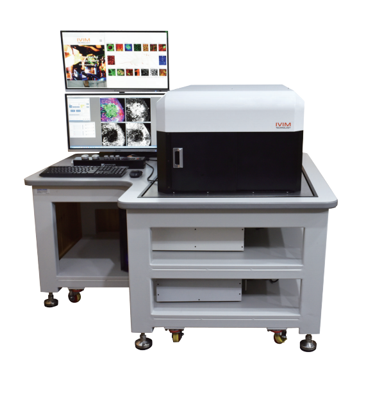
IVM-CM small animal in-situ dynamic analysis imaging system

In vivo imaging technology mainly uses a set of very sensitive optical detection equipment that can directly monitor cell activities and gene behaviors in living organisms. Through this system, biological processes such as the growth and metastasis of tum
IVM-CM technical features:
√ Wavelength range of two-photon laser: 690nm-1050nm
√ Field of view (FOV): 100*100um²-10*10mm²
√ Imaging pixels: 100fps@512*512pixels
√Imaging depth: 1000um
√In vivo imaging types: liver, lymph nodes, spleen, skin, retina, lung, brain, colon, pancreas, small intestine, prostate, kidney, heart, trachea, esophagus, esophagus, bone marrow, thymus, etc.
IVIM Technology's integrated intravital microscope system is equipped with an ultra-fast rotating polygon mirror scanner to support ultra-high-speed intravital imaging (maximum 100fps@512x512 pixels) to achieve uniform excitation and illumination over the entire field of view (FOV). No fluorescence signals and no fluorescence signals are seen in the central area of the FOV. The signal-to-noise ratio (SNR) reduces the edges of the FOV without excessive photobleaching. A uniform high signal-to-noise ratio across the FOV does not require too many photons to improve image quality.
Application areas:
●Immunology research
●Cancer research
●Targeted research
●Molecular Pathology
Application case
A new method for time-lapse imaging of immune cells in the heart of mice with myocardial infarction:
IVM-CM uses a special speculum combined with a small incision through the intercostal space to display fluorescently labeled cells and blood vessels in the heart tissue. Presents a miniature suction assisted endoscope suitable for imaging cellular events in a rat with a beating heart. The endoscope uses a suction tube device that can locally stabilize the underlying tissue without causing harmful effects, and allows imaging at the cellular level. The endoscope can enter the outer surface of the heart by making a small incision between the skin and the muscles between the ribs without breaking the bones.
IVM-CM technical parameters:
1. It is required to have the function to realize dynamic real-time imaging of the dynamic changes of various cell levels in the living body
2. In vivo imaging types: liver, lymph nodes, spleen, skin, retina, lung, brain, colon, pancreas, small intestine, prostate, kidney, heart, trachea, esophagus, esophagus, bone marrow, thymus, etc.
3. Two-photon laser wavelength range: 690nm-1050nm, pulse width <75fs, frequency range: 80MHz
4. Average power: >2.5W. Dispersion compensation: 0to-49,000fs
5. Scan head: polygon prism (fast axis scanning, maximum 66kHz); galvanometer scanner (slow axis scanning, maximum 200us/step)
6. Objective lens: optional up to 6 objective lens options (1X---100X) compatible with commercial objective lenses (RMS or M25)
7. Field of view (FOV): 100*100um²-10*10mm²
8. Imaging pixels: 100fps@512*512pixels
因用于机器人各方面应用且与大多数机器人类型兼容,AutoCal系统可以检测出机器人自身构造和工具中心点(TCP)的 突然改变或偏离,并且该系统无需人为干涉就自动地更正这些误差。
AutoCal系统-Dynalog的先进水平校准技术,Dynalog是机器人单元标定技术的世界领导者。它的主流产品DynaCal 系统,被应用于离线的机器人单元校准,并作为最精确的和技术先进的机器人校准程序为许多机器人制造商和终端使用者所接受。AutoCal 系统将已证实的DynaCal校准技术结合到一个在线的全自动系统中,该系统专为程序控制和复原而设计的,价格低廉。
AutoCal系统提供在线的机器人校准方案,旨在快速和自动地保证机械设备的工作性能。因用于机器人各方面应用且与大多数机器人类型兼容,AutoCal系统可以检测出机器人自身构造和工具中心点(TCP)的 突然改变或偏离,并且该系统无需人为干涉就自动地更正这些误差。这意味着不用猜测哪里会出错,不用浪费宝贵时间在机器人程序重复校准上,产品品质无任何损失。
AutoCal系统-Dynalog的先进水平校准技术,Dynalog是机器人单元标定技术的世界领导者。它的主流产品DynaCal 系统,被应用于离线的机器人单元校准,并作为最精确的和技术先进的机器人校准程序为许多机器人制造商和终端使用者所接受。AutoCal 系统将已证实的DynaCal校准技术结合到一个在线的全自动系统中,该系统专为程序控制和复原而设计的,价格低廉。