上海埃飞科技
Worldwide Technology(S. H)上海埃飞科技
Worldwide Technology(S. H)产品展示
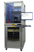

Bio-Quantum Sensor

Long-term oxidative stress can damage cells, proteins, and DNA, accelerating aging and leading to various health conditions. Real-time monitoring of cellular (or organismal) responses to oxidative stress is a challenge. Addressing these challenges, this t
Quantum Nuova is an advanced quantum sensor meticulously designed for life science research and co-developed with experts from the University Medical Center Groningen (UMCG) in the Netherlands. It enables real-time monitoring of chemical reactions at the nanomolar level and is suitable for the smallest cellular and tissue spaces. Leveraging our innovations in diamond magnetometry/gravity sensing, Quantum Nuova is finely tuned to explore sub-nanometer environments in living organisms, providing unprecedented precision and insight, making it a powerful tool in biomedical research.
Diamond magnetometry is an innovative quantum technology capable of detecting the magnetic resonance signals of free radicals in biological samples with unmatched precision. This method utilizes fluorescent nanodiamonds containing nitrogen-vacancy (NV) centers—defects in the diamond crystal structure that are highly sensitive to magnetic noise. Unlike traditional magnetic resonance methods that require complex and expensive equipment, diamond magnetometry allows the use of simple optical microscopes to capture these signals. This is made possible by the unique properties of NV centers, which convert magnetic interactions into detectable optical signals. This breakthrough technology offers a more accessible, efficient, and non-invasive method for real-time study of free radicals, making it a powerful tool in biological research and diagnostics.

Figure 1. Quantum Nuova.
Application Fields:
Healthcare, Pharmaceuticals, and Medical Diagnostics
Free radical detection in primary human granulocytes: A cutting-edge method used in fertility and reproductive health, providing deep insight into cellular stress and vitality.
Quantum detection of free radicals in dendritic cells: Crucial for advancing research in immune responses and oxidative stress-driven diseases, with potential applications in inflammatory diseases.
SARS-CoV-2 detection based on nanodiamonds: Revolutionizing diagnostic accuracy through highly sensitive quantum sensing.
Mechanism of action study of indomethacin: Utilizing quantum sensing for precise pharmacological studies to accelerate drug development.
Biotechnology and Cell Research
Quantum sensing in yeast cells: Enables real-time monitoring of cell metabolism in bioreactor cultures, driving industrial fermentation and bioengineering.
Free radical detection in bacteria: Promotes a deeper understanding of bacterial stress responses, especially in antibiotic resistance research and therapy optimization.
Materials Science and Nanotechnology
Monitoring mechanical performance and degradation of biomedical scaffolds: Enhances tissue engineering and regenerative medicine by ensuring structural integrity and durability of biomedical scaffolds.
The existing optical system includes a confocal microscope. Quantum Nuova is equipped with a 561 nm Nd:YAG laser for excitation of nitrogen-vacancy (NV) centers. The laser emits pulsed light at different intervals, with a continuous illumination power of approximately 50 µW. Emission photons from the NV centers are detected by a state-of-the-art single-photon avalanche diode.

Figure 2. First part of the optical setup.
As shown in Figure 2, the laser is guided along the path between (3) and (5). This path includes a beam-splitter cube that directs the beam through an acousto-optic modulator, followed by a λ/4 waveplate, then reflects back through the same aperture to enable fast and precise laser pulsing. This part is integrated within the laser module. Finally, the beam is coupled into an optical fiber and directed to the second part of the setup as shown in Figure 3. Here, the laser is directed via a dichroic mirror (3), which separates the incident laser light from the red fluorescence emitted by the NV centers. This is essential to allow sensitive detection and avoid interference from the excitation laser's background signal.

Figure 3. Second part of the optical setup.
Nanodiamonds are excited by the 561 nm laser. After excitation, the NV centers emit red-shifted fluorescence, which is guided back along the same path until reaching the dichroic mirror. This mirror separates the reflected green light from the red fluorescence and directs the fluorescence into the detection path (3)-(10). After passing through a pinhole, the light is detected by a single-photon-sensitive avalanche photodiode—currently the most sensitive detector for such measurements. To reduce counts from reflected light, the detection path is housed within a light-tight transportable box. The dimensions of the optical path are currently determined by the size of the lenses. Realignment of the device is fully automated (computer-controlled).
On T1 Relaxometry
T1 measurement using nitrogen-vacancy (NV) centers in nanodiamonds relies on detecting the relaxation time in response to external magnetic noise. NV centers are defects in the diamond lattice with unique spin properties that can be optically controlled and read via fluorescence. T1, or longitudinal relaxation time, indicates the time it takes for the NV spin state to return to thermal equilibrium after laser polarization. During this period, interactions with the surrounding environment—such as magnetic noise or spin-lattice interactions—cause the NV center to lose its polarization and return to the ground state. The T1 time represents the characteristic time of this process and is sensitive to magnetic field fluctuations, making it highly useful for detecting weak magnetic noise and local magnetic environments, particularly in applications for free radical measurement in living organisms.
The process of measuring T1 in NV nanodiamonds typically involves the following steps:
1. Optical Polarization: The NV centers are initially polarized using green laser light, aligning the NV spin state into a well-defined quantum state. (Figure 4a)
2. Delay Period: After laser excitation, the system is allowed to relax over a variable period. During this delay, the NV spin state interacts with its environment and starts to lose polarization. (Figure 4b)
3. Fluorescence Readout: At different delay times, the NV center fluorescence is measured. The fluorescence intensity correlates with the spin state—more polarization results in brighter fluorescence.
4. Data Fitting: Fluorescence signals at various delay times are fitted to an exponential decay model to determine the T1 relaxation time.

Figure 4.
(a) Excited state of an electron.
(b) Data acquisition and processing of relaxometry data.
(c) To perform a relaxometry experiment, the NV centers must be excited into the ms = 0 state of the excited state and then probed with a subsequent pulse to determine whether the electron decayed back to the ground state in the ms = 0 or ms = ±1 states. The more NV centers in the ms = ±1 states, the lower the fluorescence intensity (in red) in the first microsecond of each pulse. (c) To determine the magnetic noise, this intensity is plotted against the dark time and the data is fitted to generate a relaxation or T1 curve.
Software
Quantum Pulse automates the control system and data analysis in Quantum Nuova. It allows users to control laser intensity, sample stage position, nanodiamond/cell selection, brightfield/confocal imaging, physiological conditions of cells, and photon detection. Additionally, the software’s analysis functions enable measurement of T1 or spin-lattice relaxation time. Quantum Pulse is compatible with Windows OS and comes pre-installed as part of the standard Quantum Nuova package.
Key Features:
Simultaneous acquisition of brightfield and confocal images for a more comprehensive sample view.
Mosaic Imaging: Automatically stitches multiple smaller images into a large, high-resolution composite, allowing users to explore detailed regions at various scales.
Motorized Sample Stage Control: Enables automatic localization of NV centers in nanodiamonds within biological samples, achieving high-sensitivity free radical detection in live cells and tissues.
Accurate Spin-Lattice Relaxation Time Capture: Real-time tracking of free radical concentration changes in detected nanoparticles as part of standard T1 relaxometry.
Advanced Curve Fitting Algorithms: Built-in sophisticated curve fitting algorithms provide analytical functions to extract insights from direct cellular T1 measurements.

Figure 5. Quantum Pulse UI, including rightfield and confocal images (top left), T1 curve (bottom left), and operating menus (right).

因用于机器人各方面应用且与大多数机器人类型兼容,AutoCal系统可以检测出机器人自身构造和工具中心点(TCP)的 突然改变或偏离,并且该系统无需人为干涉就自动地更正这些误差。
AutoCal系统-Dynalog的先进水平校准技术,Dynalog是机器人单元标定技术的世界领导者。它的主流产品DynaCal 系统,被应用于离线的机器人单元校准,并作为最精确的和技术先进的机器人校准程序为许多机器人制造商和终端使用者所接受。AutoCal 系统将已证实的DynaCal校准技术结合到一个在线的全自动系统中,该系统专为程序控制和复原而设计的,价格低廉。
AutoCal系统提供在线的机器人校准方案,旨在快速和自动地保证机械设备的工作性能。因用于机器人各方面应用且与大多数机器人类型兼容,AutoCal系统可以检测出机器人自身构造和工具中心点(TCP)的 突然改变或偏离,并且该系统无需人为干涉就自动地更正这些误差。这意味着不用猜测哪里会出错,不用浪费宝贵时间在机器人程序重复校准上,产品品质无任何损失。
AutoCal系统-Dynalog的先进水平校准技术,Dynalog是机器人单元标定技术的世界领导者。它的主流产品DynaCal 系统,被应用于离线的机器人单元校准,并作为最精确的和技术先进的机器人校准程序为许多机器人制造商和终端使用者所接受。AutoCal 系统将已证实的DynaCal校准技术结合到一个在线的全自动系统中,该系统专为程序控制和复原而设计的,价格低廉。